Ceroid histiocytes, otherwise referred to as SBHs !! The Sea Blue Histiocytes

Ceroid histiocytes, otherwise referred to as SBHs, were first characterized by Moeschlin in 1951. Ceroid, which means “wax-like,” was first introduced as a descriptive term for a yellow-brown pigment accumulating in the cirrhotic livers of rats maintained on a low-protein, low-fat diet. Subsequently, Sawitsky et al and Silverstein et al coined the term “sea-blue histiocyte” (SBH), referring to the characteristic staining quality of ceroid with Romanowsky’s stains.

Content-16-1471-1471_full

IMAGE SOURCE : AMERICAN SOCIETY OF HEMATOLGY , ASH IMAGE BANK

Accumulation of these cells are seen a wide variety of conditions and are non specific finding in tissue sections from bone marrow of spleen. It can be seen in conditions ranging from lysosomal storage diseases to hematologic neoplastic proliferations. Common to all of these entities is an underlying abnormality in lipid metabolism, which may be due to an enzyme deficiency, as in Gaucher’s disease and Tay-Sachs disease, or an overwhelming accumulation of phospholipid-rich membranes that exceeds the maximum catabolic rate of the individual.

The histochemical staining properties of ceroid histiocytosis

[1] Wright’s and Giemsa staining imparts a sea-blue color, which also accounts for the descriptive term SBH.

[2] Positivity is observed by a PAS reaction both before and after diastase digestion

[3] By autofluorescence and acid-fast fluorescence, with reactions tending to increase with aging of the slide

[4] Sudan black stain.

NOTE :- Ceroid does not react with Kinyoun carbolfuchsin or Prussian blue stains or in using Fite’s method.

FOR MORE THAN 300 IMAGES BASED Q VISIT pgmeeuploads.com

Dengue TESTS : What to do and when ?

Dengue can be diagnosed by isolation of the virus, by serological tests, or by molecular methods. Diagnosis of acute (on-going) or recent dengue infection can be established by testing serum samples during the first 5 days of symptoms and/or early convalescent phase (more than 5 days of symptoms).

Before day 5 of illness, during the febrile period, dengue infections may be diagnosed by virus isolation in cell culture, by detection of viral RNA by nucleic acid amplification tests (NAAT), or by detection of viral antigens by ELISA or rapid tests.

NOTE 1: The non-structural protein 1 (NS1) of the dengue viral genome is a useful as a tool for the diagnosis of acute dengue infections. Dengue NS1 antigen has been detected in the serum of infected patients as early as 1 day post onset of symptoms, and up to 7.  The NS1 assay may also be useful for differential diagnostics between flaviviruses because of the specificity of the assay.

NOTE 2 : After day 5, dengue viruses and antigens disappear from the blood coincident with the appearance of specific antibodies. NS1 antigen may be detected in some patients for a few days after defervescence.

NOTE 3 : Plaque Reduction and Neutralization Test (PRNT) and the microneutralization PRNT can be used when a serological specific diagnostic is required, as this assay is the most specific serological tool for the determination of dengue antibodies

Following table shows the utility of tests ( WHO 2009)

Diagnostic methods Diagnosis of acute infection Time to results Specimen Time of collection after onset of symptoms
Viral isolation and serotype identification Confirmed 1–2 weeks Whole blood, serum, tissues 1–5 days
Nucleic acid detection Confirmed 1 or 2 days Tissues, whole blood, serum, plasma 1–5 days
Antigen detection Not yet determined 1 day Serum 1–6 days
Confirmed >1 day Tissue for immuno-chemistry NA
IgM ELISA Probable 1–2 days Serum, plasma, whole blood After 5 days
IgM rapid test 30 minutes
IgG (paired sera) by ELISA, HI or neutralization test Confirmed 7 days or more Serum, plasma, whole blood Acute sera, 1–5 days; convalescent after 15 days

FDP vs D DIMER : THE GAMES OF SENSITIVITY AND SPECIFICITY

FDP vs D DIMER

First we need to understand clot formation with this perspective

CLOT FORMATION

First platelet plug is formed than fibrin (from a bigger molecule called fibrinogen) sticks to the platelet plug and seals it up. When fibrin seals up a clot, there’s actually an extra, final step in which factor XIII creates little cross-links between the fibrin molecules.

NOW we need to Know how FDP and D DIMERs are produced ?

Plasmin can attack the fibrin in the plug, chopping it up into little fragments called fibrin degradation products, which can BE measured in the blood. This breakdown process actually starts happening almost right away, as soon a clot is being formed.

What is the difference between FDPs and D-dimers?

Fibrin degradation products (FDPs) and D-dimers are both little fibrin-containing molecules formed by fibrin degredation. D-dimers are different from FDP as  they contain an extra little linkage. When fibrin seals up a clot, there’s actually an extra, final step in which factor XIII creates little cross-links between the fibrin molecules. When the fibrin degredation occurs, some of the resulting fragments will contain these cross-links and are called D-dimers.

SO D DIMERS ARE ESSENTIALLY A TYPE OF FDP

SO FDP IS AN UMBRELLA UNDER WHICH D DIMER COMES

SO  TESTING FOR FDP

[1] Test is SUPER SUPER sensitive. Meaning that even small, normal clots give off enough FDPs to make the FDP test register as positive.

[2] The second problem is that FDPs can be created from circulating fibrinogen too. So if you see FDPs, you don’t really know if they came from a clot or not and hense NON SPECIFIC

D-dimers are more specific for actual clots than FDPs are – because you only get D-dimers from the breakdown of real clots AND not from the breakdown of fibrinogen.

Tests that specifically look for D-dimers were developed in the 1990s, and most labs use these D-dimer assays now instead of assays that measure FDPs. However it still has high sensitivity andlow specificity so it cant be confirmatory to rule in a thrombus in presence of D DIMER

Best use of the D-dimer assay = So it’s great for ruling out a thrombus.

SO FOR DIC : MOST SENSITIVE = FDP , MOST SPECIFIC = D DIMER

NORMAL SKIN HISTOLOGY

The skin or cutis covers the entire outer surface of the body. Structurally, the skin consists of two layers which differ in function, histological appearance and their embryological origin. The outer layer or epidermis is formed by an epithelium and is of ectodermal origin. The underlying thicker layer, the dermis, consists of connective tissue and develops from the mesoderm. Beneath the two layers we find a subcutaneous layer of loose connective tissue, the hypodermis or subcutis, which binds the skin to underlying structures. Hair, nails and sweat and sebaceous glands are of epithelial origin and collectively called the appendages of the skin.

The epidermis consists of four clearly defined layers or strata:

• Basal cell layer (stratum basale)
• Prickle cell layer (stratum spinosum)
• Granular cell layer (stratum granulosum)
• Keratin layer (stratum corneum)

NOTE : An eosinophilic acellular layer known as the stratum lucidum is seen in skin from the palms and soles.

histology-skin-part-1-23-728

The stratum basale = is the deepest layer of the epidermis. It consists of a single layer of  cuboidal or columnar cells with a large nucleus typically containing a conspicuous nucleolus. Small numbers of mitoses may be evident.Their mitotic activity replenishes the cells in more superficial layers as these are eventually shed from the epidermis. The renewal of the human epidermis takes about 3 to 4 weeks.

The stratum spinosum = Histologically, prickle cells are polygonal in outline, have abundant eosinophilic cytoplasm and oval vesicular nuclei, often with conspicuous nucleoli. The cells are often separated by narrow, translucent clefts. These clefts are spanned by spine-like cytoplasmatic extensions of the cells (hence the name of the layer and of its cells: spinous cells), which interconnect the cells of this layer. Spines of cells meet end-to-end or side-to-side and are attached to each other by desmosomes.

NOTE : In addition to the usual organelles of cells, EM shows membrane-bound lamellar granules in the cytoplasm of the spinous cells.

Clear cells are present in the basal layer of the epidermis = Melanocytes.

NOTE :

(1) Cells with clear cytoplasm seen in the stratum spinosum represent Langerhans cells = the epidermis also contains a network of about 2 × 10⁹ Langerhans cells which serve as sentinel cells whose prime function is to initiate immune responses against microbial threats.

(2) Toker cells represent an additional clear cell population, which may be found in nipple epidermis of both sexes in up to 10% of the population. The  cells are large, polygonal or oval and have abundant pale staining or clear
cytoplasm with vesicular nuclei often containing prominent, albeit small, nucleoli. the cytoplasm is mucicarmine and PAS negative. Toker cells may be mistaken as paget cells.

The stratum granulosum = consists, in thick skin, of a few layers of flattened cells. Only one layer may be visible in thin skin. The cytoplasm of the cells contains numerous fine grains, keratohyalin granules. Keratohyalin granules typify the granular cell layer . The keratohyalin is not located in membrane-bound organelles but forms “free” accumulations in the cytoplasm of the cells.

The stratum lucidum = consists of several layers of flattened dead cells. Nuclei already begin to degenerate in the outer part of the stratum granulosum. In the stratum lucidum, faint nuclear outlines are visible in only a few of the cells. The stratum lucidum can usually not be identified in thin skin.

NOTE : An eosinophilic acellular layer known as the stratum lucidum is seen in skin from the palms and soles.

The stratum corneum = cells are completely filled with keratin filaments (horny cells) which are embedded in a dense matrix of proteins. The protection of the body by the epidermis is essentially due to the functional features of the stratum corneum.

NOTE :  The cornified cell envelope and the stratum corneum restrict water loss from the skin while keratinocyte-derived endogenous antibiotics (defensins and cathelicidins) provide an innate immune defense against bacteria, viruses and fungi.

Eccrine sweat glands are found at all skin sites and are present in densities of 100–600/cm2; they play a role in heat control and aspects of metabolism. Secretions from apocrine sweat glands contribute to body odor. Skin lubrication and waterproofing is provided by sebum secreted from sebaceous glands.

Sweat Glands

Two types of sweat glands are present in humans.

1. Merocrine (~eccrine) sweat gland

The skin contains ~3,000,000 sweat gland which are found all over the body – with the exception of, parts of the external genitalia. Sweat glands are simple tubular glands. The secretory epithelium is cuboidal or low columnar. Two types of cells may be distinguished: a light type, which secretes the watery eccrine sweat, and a dark type, which may produce a mucin-like secretion. The cells have slightly different shapes and, as a result of the different shapes, the epithelium may appear pseudostratified. A layer of myoepithelial cells is found between the secretory cells of the epithelium and the basement membrane. The excretory duct has a stratified cuboidal epithelium (two layers of cells). The excretory ducts of merocrine sweat glands empty directly onto the surface of the skin.

2. Apocrine sweat glands are stimulated by sexual hormones and are not fully developed or functional before puberty. Apocrine sweat is a milky, proteinaceous and odourless secretion. The odour is a result of bacterial decomposition and is, at least in mammals other than humans, of importance for sexual attraction. The histological structure of apocrine sweat glands is similar to that of merocrine sweat glands, but the secretory epithelium consists of only one major cell type, which looks cuboidal or low columnar. Apocrine sweat glands as such are also much larger than merocrine sweat glands.

The excretory duct of apocrine sweat glands does not open directly onto the surface of the skin. Instead, the excretory duct empties the sweat into the upper part of the hair follicle. Apocrine sweat glands are therefore part of the pilosebaceous unit.

Sebaceous Glands

Sebaceous glands empty their secretory product into the upper parts of the hair follicles. They are therefore found in parts of the skin where hair is present. The hair follicle and its associated sebaceous gland form a pilosebaceous unit. Sebaceous glands are also found in some of the areas where no hair is present, for example, lips, oral surfaces of the cheeks and external genitalia. Sebaceous glands are as a rule simple and branched.

EMBRYOLOGY POINTS

Hair follicles and nails are evident at 9 weeks. The earliest development of hair occurs at about 9 weeks in the regions of the eyebrow, upper lip and chin.

Sweat glands are also noted at 9 weeks on the palms and the soles.

Sweat glands at other sites and sebaceous glands appear at 15 weeks.

Sebaceous glands first appear as hemispherical protuberances on the posterior surfaces of the hair pegs and become differentiated at 13–15 weeks.

Langerhans cells are derived from the monocyte–macrophage–histiocyte lineage and enter the epidermis at about 12 weeks.

NOW LETS IDENTIFY DIFFERENT LAYERS IN EPIDERMIS IN IMAGE GIVEN BELOW

Screenshot (781)

A = Basal cell layer (stratum basale)
B = Prickle cell layer (stratum spinosum)
C = Granular cell layer (stratum granulosum)
D = Stratum lucidum

E = Keratin layer (stratum corneum)

NOTE = The Malpighian layer of the skin is generally defined as both the stratum basale and stratum spinosum as a unit, although it is occasionally defined as the stratum basale specifically. It is named after Marcello Malpighi.

Why does cardiac muscle does not show Tetany !!!!

Why is it important to prevent Myocardial muscle from getting tetanised ??

Prevention of tetanus in the heart is important because cardiac muscles must relax between contractions so the ventricles can fill with blood.

So how does this happen ?

The longer myocardial action potential of myocardium helps prevent the sustained contraction called tetanus.

To understand how a longer action potential prevents tetanus, relationship between action potentials, refractory periods, and contraction in skeletal and cardiac muscle cells.

Refractory period is the time following an action potential during which a normal stimulus cannot
trigger a second action potential.

In cardiac muscle, the long action potential (red curve) means the refractory period and the contraction end almost
simultaneously. By the time a second action potential can take place, the myocardial cell has almost completely
relaxed. Consequently, no summation occurs. See figure below.ihg700

In contrast, the skeletal muscle action potential and refractory period are ending just as contraction begins. For this reason, a second action potential fired immediately after the refractory period causes summation of the contractions. If a series of action potentials occurs in rapid succession, the sustained contraction known as tetanus results. See figure below.okjf

Radiation Oncogenesis

1. Which of the following statements is false?

(A) Radiation damage can cause a cell to either die or continue being viable but mutated.

(B) The two events above are very different when it comes to the dose dependence of their probability to occur and also when it comes to their impact on the organism to which the cell belongs.

(C) Cell killing by radiation is said to be a “deterministic effect”.

(D) Cell mutating by radiation is said to be a “nonstochastic effect”.

2.  A deterministic effect:

(A) Has a threshold in dose but the severity of the effect is otherwise dose-independent.

(B) Has a threshold in dose and the severity of the effect increases with dose.

(C) Has no threshold in dose and the severity of the effect is a constant function of dose.

(D) Has no threshold in dose and the severity of the effect increases with dose.

3. Which of the following statements is false?

(A) Stochastic effects have no threshold in dose.

(B) The probability of induction of a stochastic effect increases with dose.

(C) The severity of a stochastic effect increases with dose.

(D) A cancer induced by 1 Gy is not worse than a cancer induced by 0.5 Gy.

4. Carcinogenesis and hereditary effects are:

(A) Deterministc effects.

(B) Stochastic effects.

(C) Both

(D) None

Have these questions generated enough interest in you ! If yes scroll down to know the answers !!!!

Screenshot (764)

Screenshot (765)

NOW COMING TO QUESTIONS ON RADIATION ONCOGENESIS

STOCHASTIC EFFECT vs DETERMINISTIC EFFECT

From the biological effects of radiation on human body, radiation effects are generally divided into two categories: “Deterministic effects” and “Stochastic effects”.

STOCHASTIC EFFECT

Radiation oncogenesis is a stochastic effect i.e. there is no threshold point and risk increases in a linear-quadratic fashion with dose. This is known as the linear-quadratic no threshold theory. Although the risk increases with dose, the severity of the effects do not; the patient will either develop cancer or they will not.

Characteristics of stochastic effects:

  1. Severity is independent of absorbed dose
  2. Threshold does not exist
  3. Probability of occurrence depends on absorbed dose

Example: radiation induced cancer, genetic effect

DETERMINISTIC EFFECT

Based on a large number of experiments involving animals and other researches, further supplemented by theoretical studies, it was discovered that severity of certain effects on human beings will increase with increasing doses. There exists a certain level, the “threshold”, below which the effect will be absent. This kind of effects is called “deterministic effects”.

Characteristics of deterministic effects:

  1. Damage depends on absorbed dose
  2. Threshold exists

Example: cataract, erythema, infertility etc.

Severity of deterministic effects depends on dose. However, thresholds exist, only above which the effects will occur. The International Commission on Radiological Protection (ICRP) considers that if the annual radiation doses to the lens of the eyes of radiation workers are restricted to 150 mSv (equivalent to 150 mGy for X-ray), cataract is unlikely to occur during his/her life assuming a working period of 50 years. For other major organs, the annual dose limits for preventing deterministic effects are as follows.

Threshold for deterministic effects (Sv)
Effects One single absorption (Sv) Prolong absorption (Sv-year)
Testis Permanent infertility 3.5 – 6.0 2
Ovary Permanent infertility 2.5 – 6.0 > 0.2
Lens of eyes Milky of lens
cataract
0.5 – 2.0
5.0
> 0.1
> 0.15
Bone marrow Blood forming deficiency 0.5 > 0.4

SO THE ANSWERS ARE

Q1. D,  Q2. B,  Q3. C,  Q4. B

IMMUNE DEPOSITS IN RENAL DISEASE

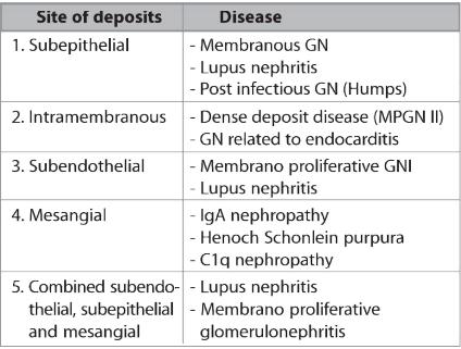
Immunofluorescence and electron microscopy are used for detection of immune deposits in case of renal biopsy.

Immunofluorescence: In this technique, as soon as renal biopsy core is obtained, it is snap frozen and cut into 2-4 µm thick sections. Later these sections are stained with antisera known to be monospecific for IgG, IgM, IgA, K & lamda light chains, C3, C1q and albumin. Then these stained sections are seen under fluorescent microscope. If deposition is granular it indicates immune complex disease and if deposition is intensely linear it indicates deposition of classic antiglomerular basement membrane antibodies.

Screenshot (759)

Electron microscopy:
For electron microscopy biopsy is fixed in gluteraldehyde or osmium tetroxide, embedded in epoxy resins and stained with toluidine blue.

Screenshot (760)

Screenshot (761)

The dying cells and how they PLAN their death !!!

Programmed cell death may be more accurately defined as cell death that is dependent on genetically encoded signals or activities within the dying cell. Apoptosis and programmed cell death are often used as synonyms. However, a variety of other molecular cell death pathways have been characterized.

APOPTOSIS = Mediated by a subset of caspases morphology includes nuclear and cytoplasmic condensation and formation of membrane-bound cellular fragments or apoptotic bodies; not inflammatory.

AUTOPHAGY = Degradation of cellular components within the dying cell in autophagic vacuoles; not inflammatory.

ONCOSIS = Prelethal pathway leading to cell death accompanied by cellular and organelle swelling and increased membrane permeability; proinflammatory.

PYROPTOSIS = Proinflammatory pathway resulting from caspase-1 activity leading to membrane breakdown and proinflammatory cytokine processing.

NECROPTOSIS = Necroptosis is a newly discovered pathway of regulated necrosis that requires the proteins RIPK3 and MLKL and is induced by death receptors, interferons, toll-like receptors, intracellular RNA and DNA sensors. This process is caspase independent and inflammatory.

zii0040547300002

IMAGE REFERENCE :
Infect Immun. 2005 Apr; 73(4): 1907–1916.

Apoptosis =  cell death = activation of initiator caspases = activate effector caspases to cleave cellular substrates. Apoptotic cells demonstrate cytoplasmic and nuclear condensation, DNA damage, formation of apoptotic bodies, maintenance of an intact plasma membrane, and exposure of surface molecules targeting intact cell corpses for phagocytosis. In the absence of phagocytosis, apoptotic bodies may proceed to lysis and secondary or apoptotic necrosis.

Autophagy features degradation of cellular components within the intact dying cell in autophagic vacuoles. The morphological characteristics of autophagy include vacuolization, degradation of cytoplasmic contents, and slight chromatin condensation. Autophagic cells can also be taken up by phagocytosis.

Oncosis is the prelethal pathway leading to cell death accompanied by cellular and organelle swelling and membrane breakdown, with the eventual release of inflammatory cellular contents.

Pyroptosis is a pathway to cell death mediated by the activation of caspase-1, a protease that also activates the inflammatory cytokines, IL-1β, and IL-18. This pathway is therefore inherently proinflammatory. Pyroptosis also features cell lysis and release of inflammatory cellular contents.

SO

Caspase dependent programmed cell death = Apoptosis & Pyroptosis

Caspase independent programmed cell death = Necroptosis & Oncosis